E coli under microscope 1000x 852240-E coli under microscope 1000x
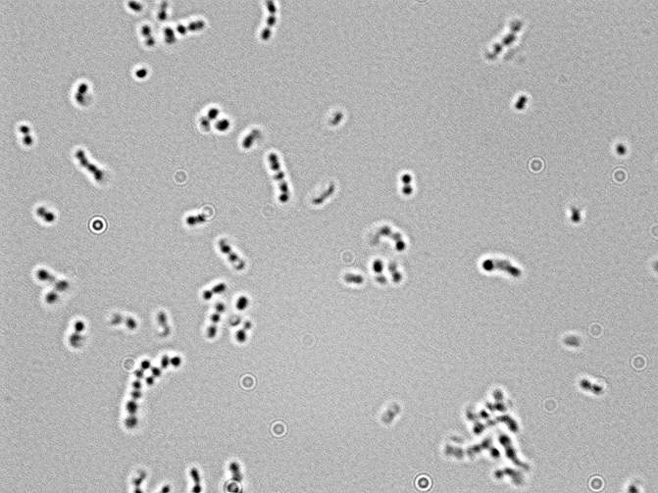
Escherichia coli bacteria on blood agar e coli under a microscope stock pictures, royaltyfree photos & images petri dishes with culture media for sarscov2 diagnostics, test coronavirus covid19, microbiological analysis e coli under a microscope stock pictures, royaltyfree photos & imagesNotice that the cells appear much smaller than in the image that has Ecoli with green color added This image was generated using a light microscope which typically magnifies Jennifer Julizar BIOL 351 Section 906 Staining Results and Conclusions using microscope under 1000X magnification with oil immersionAs indicated in the student notes, the first slide that was prepared using aseptic technique and the Gram Staining method was a slide containing cultures of M luteus and E coli When examining this slide under the microscope at 1000X magnification, I observed some clusters and mostly doubles of coccishaped purple colonies
Escherichia Coli Bacteria E Coli Stock Footage Video 100 Royalty Free Shutterstock
E coli under microscope 1000x
E coli under microscope 1000x-E coli stained gram negative under 1000x oil immersionNotice that the cells appear much smaller than in the image that has Ecoli with green color added This image was generated using a light microscope which typically magnifies Jennifer Julizar BIOL 351 Section 906 Staining Results and Conclusions using microscope under 1000X magnification with oil immersion
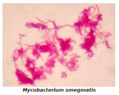


Microbiology Lab Exercise 6 Acid Fast Staining Flashcards Cram Com
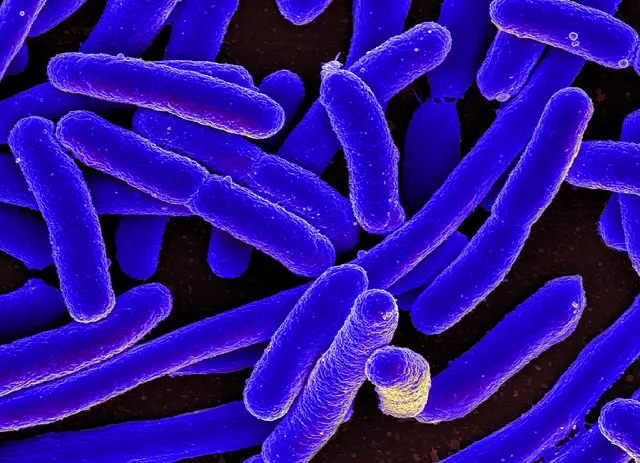
Escherichia coli (/ ˌ ɛ ʃ ə ˈ r ɪ k i ə ˈ k oʊ l aɪ /), also known as E coli (/ ˌ iː ˈ k oʊ l aɪ /), is a Gramnegative, facultative anaerobic, rodshaped, coliform bacterium of the genus Escherichia that is commonly found in the lower intestine of warmblooded organisms (endotherms) Most E coli strains are harmless, but some serotypes (EPEC, ETEC etc) can cause serious foodMixed Culture (Gram Stain) E coli and S epidermidis (1000X) Klebsiella pneumoniae (1000X) Capsule Stain Capsule Stain (1000X) Commercially Prepared Slide In some bacteria, the cell wall is surrounded by a viscous cell envelope called 'capsule' It is made of polysaccharide, glycoprotein or polypeptideThe length can range from 110 µm for filamentous or rodshaped bacteria The most wellknown bacteria E coli, their average size is ~15 µm in diameter and 26 µm in length In this figure The size comparison between our hair (~ 60 µm) and E coli (~1 µm) Notice how small the bacteria are It requires 1000x magnification to see them
Instead, their genetic material floats uncovered4 Applying counterstain (safrinin) to bacterial smear as last step of endospore stain;When viewed under the microscope, Gramnegative E Coli will appear pink in color The absence of this (of purple color) is indicative of Grampositive bacteria and the absence of Gramnegative E Coli Escherichia coli under 10х90х magnification using fuchsine as a dye by ElNokko (Own work) CC BYSA 40 (http//creativecommonsorg/licenses/bysa/40), via Wikimedia Commons
Biology Of E Coli E coli (Escherichia coli) are a small, Gramnegative species of bacteriaMost strains of E coli are rodshaped and measure about μm long and 0210 μm in diameterThey typically have a cell volume of 0607 μm, most of which is filled by the cytoplasm Since it is a prokaryote, E coli don't have nuclei;Bacteria species E coli and S aureus under the microscope with different magnifications Bacteria are among the smallest, simplest and most ancient livingOscillatoria is about 7 µm in diameter The bacterium, Epulosiscium fishelsoni , can be seen with the naked eye (600 µm long by 80 µm in diameter)
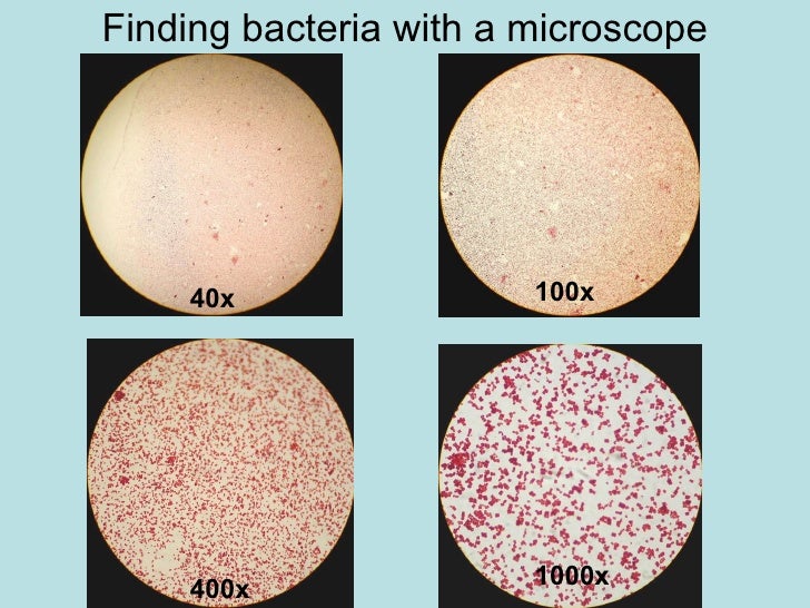


Chapter 3 Tools Of The Laboratory E Mail


Nc Dna Day A Portrait Of A Protein
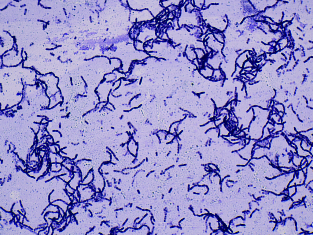
When properly Gramstained, Kocuria rhizophilia appears as purple spheres (Gram cocci) and Bacillus subtilis appears as light red rods (Gram– bacilli) at X magnification These two bacteria will be used as positive controls for comparison when Gramstaining a sample of E coliE Coli (Escherichia Coli) is a gramnegative, rodshaped bacterium Most E Coli strains are harmless, but some serotypes can cause food poisoning in their hosts The harmless strains are part of the normal flora of the gut Learn more about E Coli here Helicobacter Pylori The prepared microscope slide image of Helicobacter Pylori at left was captured at 400x magnificationEscherichia coli (/ ˌ ɛ ʃ ə ˈ r ɪ k i ə ˈ k oʊ l aɪ /), also known as E coli (/ ˌ iː ˈ k oʊ l aɪ /), is a Gramnegative, facultative anaerobic, rodshaped, coliform bacterium of the genus Escherichia that is commonly found in the lower intestine of warmblooded organisms (endotherms) Most E coli strains are harmless, but some serotypes (EPEC, ETEC etc) can cause serious food


In Vitro Study Of The Activity Of Some Medicinal Plant Leaf Extracts On Urinary Tract Infection Causing Bacterial Pathogens Isolated From Indigenous People Of Bolangir District Odisha India Biorxiv


Bacteria Under The Microscope E Coli And S Aureus Youtube
Field of view can be measured by using the 100X ruler(a 'student' millimeter ruler as seen under 100 power magnification) and counting and estimating the number of mm's in the view at the widest point, the diameter (app 15 mm here, individual microscopes will vary) Ignore the 400X circle for the moment3 Malachite green primary staining step of endopore stain with slide being heated over water bath;Focusing With The 1000X Oil Immersion Objective Olympus CX31 Microscope Place a rounded drop of immersion oil on the area of the slide that is to be observed under the microscope, Escherichia coli is a small bacillus Estimate the length and width of a typical rod c


E Coli Bacteria Magnified 12 000x Vettechlife Electron Microscope Images Electron Microscope Microscopic Images


What Does An E Coli Bacteria Look Like Under A Microscope Quora
2 Negative endospore stain showing only vegetative cells @1000xTM;Focusing With The 1000X Oil Immersion Objective Olympus CX31 Microscope Place a rounded drop of immersion oil on the area of the slide that is to be observed under the microscope, Escherichia coli is a small bacillus Estimate the length and width of a typical rod cWhich bacteria look similar to E coli under 100X optical microscope?


Microbiology Lab Exercise 6 Acid Fast Staining Flashcards Cram Com
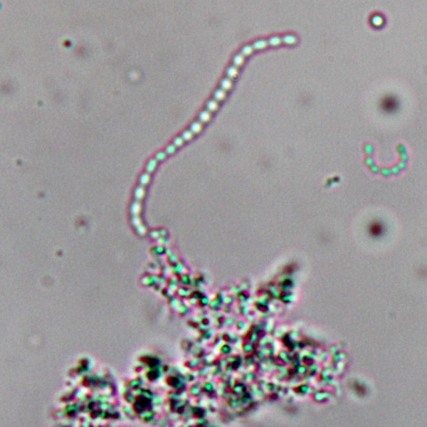


Observing Bacteria Under The Light Microscope Microbehunter Microscopy
Phase contrast microscopy was used to observe the live E coli and B megaterium cells on wet mount slides under 1000x magnification The E coli cell sizes ranged from 1 µm 6 µm, but most of them averaged around 2 µm 3 µm The E coli cells were bacilli shaped and uniform throughout the sample The E coli cellsEscherichia coli, often abbreviated E coli, are rodshaped bacteria that tend to occur individually and in large clumps E coli are classified as facultative anaerobes, which means that they grow best when oxygen is present but are able to switch to nonoxygendependent chemical processes in the absence of oxygenPlace the slide on a staining rack and add a few drops of crystal violet onto the sample, gently wash with water Add a few drops of Gram iodine (mordant) for between 30 seconds and 1 minute, gently wash with water Add a few drops of alcohol (95% alcohol) for about 10 seconds, gently wash with water
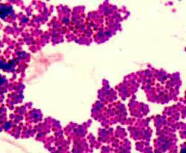


Bacterial Staining Microbiology Images Photographs And Videos Of Gram Acid Fast Endospore


Microscope World Blog Microscopy Gram Staining
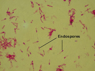
Endospore stained slide, with control Bacillus on left, negative control E coli on right, and unkown in centerEndospore stained slide, with2 Negative endospore stain showing only vegetative cells @1000xTM;
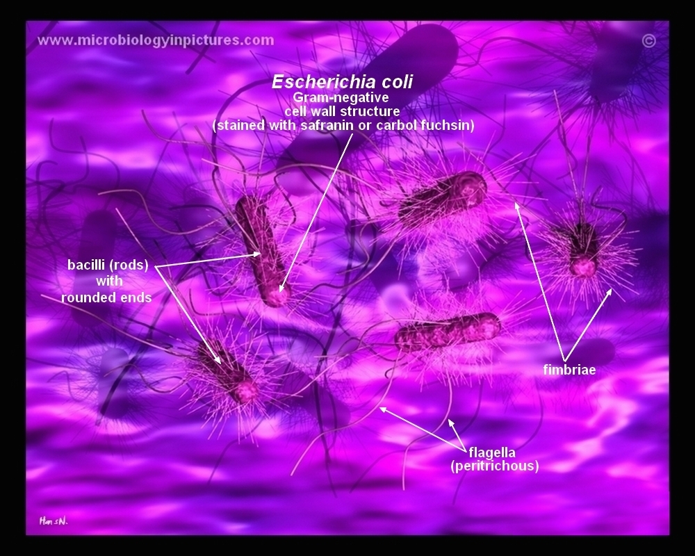


How E Coli Bacteria Look Like


Escherichia Coli Bacteria E Coli Stock Footage Video 100 Royalty Free Shutterstock
E coli under the microscope Escherichia coli (E coli) is a bacterium commonly found in various ecosystems like land and water Most of the strains of E coli are harmless, but some strains are known to cause diarrhea and even UTIs E coli is commonly studied as they are considered as a standard for the study of different bacteriaEscherichia coli or Ecoli is a gramnegative species of bacillus shaped bacteria that can be easily observed under a microscope, even for those with the untrained eye This bacteria has a fast growth rate, doubling every minutes, making them a common choice for bacterial related research purposesLarge, beige, drylike colonies Escherichia coli Small, pinpoint or dotlike, white colonies Bacteria E coli Must observe under 400x Very small & motile Looks like specks of sand Gram Stain must view under 1000x
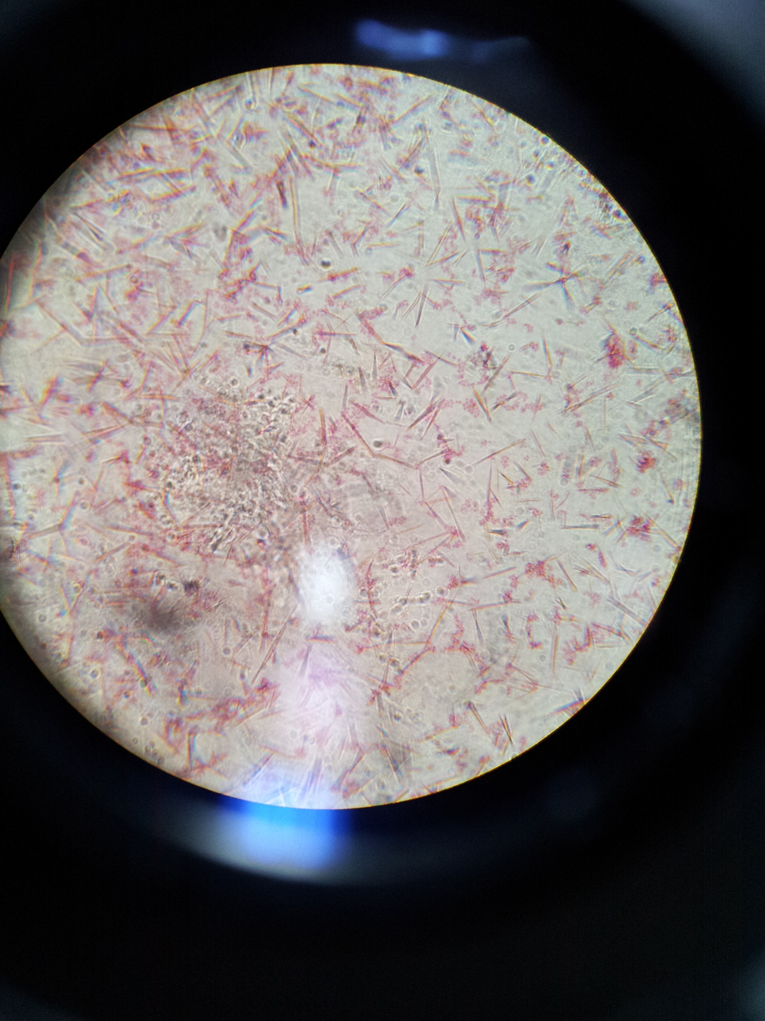


Lab 1 Principles And Use Of Microscope Ibg 102 Lab Reports


White Blood Cells Crawling Under The Microscope 1000x Magnification Youtube
Endospore stain of Bacillus subtilis showing both endospores (green) & vegetative cells (pink) @1000xTM;Although proteins in both E coli and M tuberculosis have disulfide bonds, the bonds are formed differently in each of the organisms M tuberculosis uses an enzyme called vitamin K epoxide reductase (VKOR) This enzyme, important for blood coagulation pathways in humans, is the target of the drug warfarin (commercially known as Coumadin)• A basic light microscope has 4 objective lenses – 4X, 10X, 40X, and 100X The higher the number, the higher the power of magnification Once you have focused the


Escherichia Coli Bacteria E Coli Stock Footage Video 100 Royalty Free Shutterstock


C Met At C Mould Exploring The Invisible
E coli under the microscope Escherichia coli (E coli) is a bacterium commonly found in various ecosystems like land and water Most of the strains of E coli are harmless, but some strains are known to cause diarrhea and even UTIs E coli is commonly studied as they are considered as a standard for the study of different bacteria3 Malachite green primary staining step of endopore stain with slide being heated over water bath;The resolution of a microscope depends on the wavelength of light used 3 Increasing magnification of an image will also increase the resolving power All the cells appear blue under the oil lens This is an example of Simple staining Brainheart infusion, trypticase soy agar (TSA), and nutrient agar are all examples of which type of


Gram Stain


Figure 3
Ecoli is usually motile in liquid or semisolid environment with peritrichous flagella (about 6 per cell) and its surface is covered with fimbriae These structures (flagella and fimbriae) are too thin to be visualized by classical light microscopy or they don't have to be present at all under given cultivation conditions even at motile strainsPhase contrast microscopy was used to observe the live E coli and B megaterium cells on wet mount slides under 1000x magnification The E coli cell sizes ranged from 1 µm 6 µm, but most of them averaged around 2 µm 3 µm The E coli cells were bacilli shaped and uniform throughout the sample The E coli cellsE coli under microscope 1000x E Coli Under Microscope 1000x Written By MacPride Saturday, July 21, 18 Add Comment Edit Chapter 3 Tools Of The Laboratory E Mail Balantidiasis Microscopy Findings Bacteria Bacilli And Cocci Colored With The Gram Staining Flickr Lactobacillus Acidophilus Wikipedia


Microscopy Gram Staining Microscope World Blog
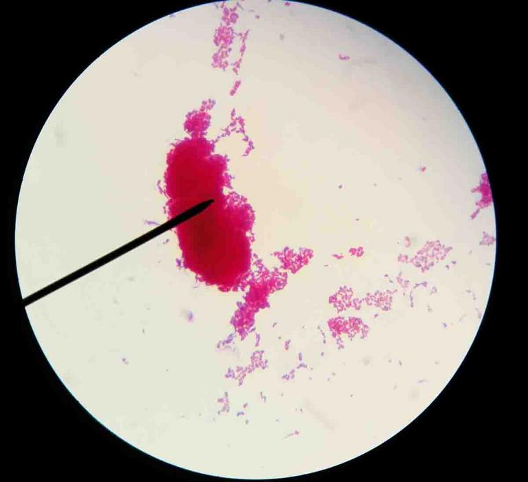


Acid Fast Stain Free Microbiology Images From Science Prof Online
Examples of Bacteria Under the Microscope Escherichia coli Escherichia coli (Ecoli) is a common gramnegative bacterial species that is often one of the first ones to be observed by students Most strains of Ecoli are harmless to humans, but some are pathogens and are responsible for gastrointestinal infections They are a bacillus shapedSalmonella includes a group of gramnegative bacillus bacteria that causes food poisoning and the consequent infection of the intestinal tract While some of the infections can be easily treated, some of the strains have been shown to resist antibiotic treatmentE Coli E Coli under the microscope at 400x E Coli (Escherichia Coli) is a gramnegative, rodshaped bacterium Most E Coli strains are harmless, but some serotypes can cause food poisoning in their hosts The harmless strains are part of the normal flora of the gut
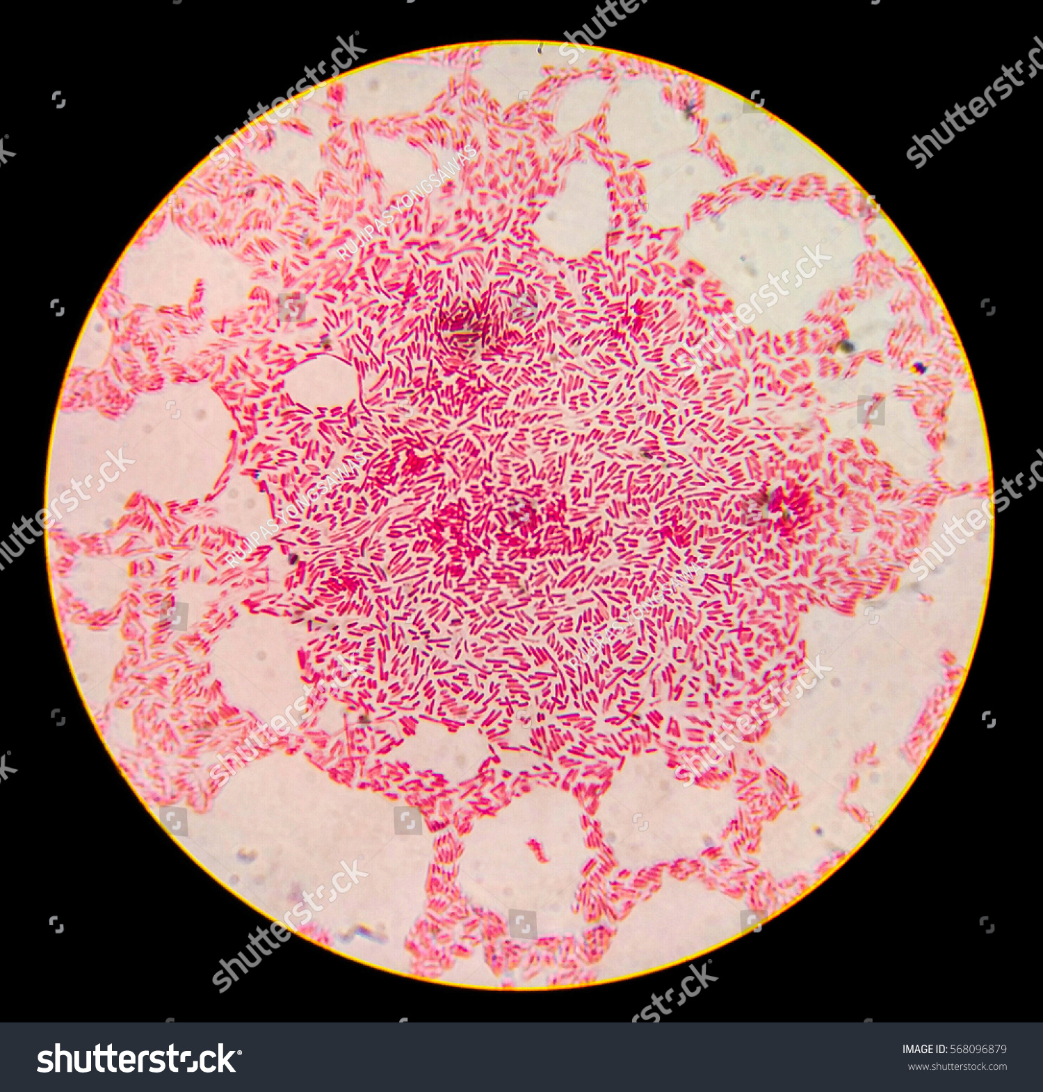


Escherichia Coli Gram Staining Compound Microscope Stock Photo Edit Now


Gram Stain
4 Applying counterstain (safrinin) to bacterial smear as last step of endospore stain;2 Morphology and Staining of Escherichia Coli E coli is Gramnegative straight rod, 13 µ x 0407 µ, arranged singly or in pairs (Fig 281) It is motile by peritrichous flagellae, though some strains are nonmotile Spores are not formed Capsules and fimbriae are found in some strains 3 Cultural Characteristics of Escherichia ColiE coli under microscope 1000x › e coli under microscope 100x › e coli under microscope 10x › e coli under e coli under microscope oil immersion E Coli Under Microscope Written By MacPride Friday, June 14, 19 Add Comment Edit British E Coli Cases Rise World News The Guardian Microscopic Studies Of Various Organisms


Www Mccc Edu Hilkerd Documents Bio1lab3 Exp 4 Pdf


Morphology Of E Coli Cells Under Microscope At 100 Magnification Download Scientific Diagram
Field of view can be measured by using the 100X ruler(a 'student' millimeter ruler as seen under 100 power magnification) and counting and estimating the number of mm's in the view at the widest point, the diameter (app 15 mm here, individual microscopes will vary) Ignore the 400X circle for the momentResearcher with e coli bacteria bacteria under microscope stock pictures, royaltyfree photos & images ebola outbreak infographic bacteria under microscope stock illustrations chemical test of water bacteria under microscope stock pictures, royaltyfree photos & imagesEscherichia coli light microscopy Escherichia coli Magnification 1000×


Gram Stain
/gram-positive-staphylococcus-aureus-bacteria-541802136-57979cca5f9b58461f26eccc.jpg)


Gram Stain Procedure In Microbiology
The easiest way to see bacteria through a microscope is to prepare a bacterial smear A smear is a bacterial sample mixed into a drop of water on a microscope slide and then heat fixed By placing the slide on a microincinerator tray or gently waving slide over Bunsen burner flame until dryE coli , a bacillus of about average size is 11 to 15 µm wide by to 60 µm long Spirochaetes occasionally reach 500 µm in length and the cyanobacterium;As indicated in the student notes, the first slide that was prepared using aseptic technique and the Gram Staining method was a slide containing cultures of M luteus and E coli When examining this slide under the microscope at 1000X magnification, I observed some clusters and mostly doubles of coccishaped purple colonies


What Does An E Coli Bacteria Look Like Under A Microscope Quora


Escherichia Coli Light Microscopy
1 E coli stained with crystal violet @ 100xTM It is not possible to see individual bacteria at this magnification, but viewing at 100XTM is a necessary step in focusing to scope to see at higher magnifications 2 E coli stained with crystal violet @ 1000xTMEndospores are formed by a few genera of bacteria, such as Bacillus By forming spores, bacteria can survive in hostile conditions Spores are resistant tExamples of Bacteria Under the Microscope Escherichia coli Escherichia coli (Ecoli) is a common gramnegative bacterial species that is often one of the first ones to be observed by students Most strains of Ecoli are harmless to humans, but some are pathogens and are responsible for gastrointestinal infections They are a bacillus shaped
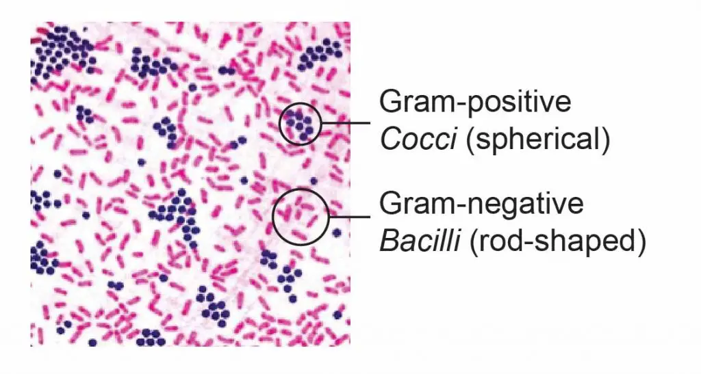


Observing Bacteria Under The Microscope Gram Stain Steps Rs Science


Biol 230 Lab Manual Lab 1
Escherichia coli light microscopy Escherichia coli Magnification 1000×1 Endospore stain of Bacillus subtilis showing both endospores (green) & vegetative cells (pink) @1000xTM;The length can range from 110 µm for filamentous or rodshaped bacteria The most wellknown bacteria E coli, their average size is ~15 µm in diameter and 26 µm in length In this figure The size comparison between our hair (~ 60 µm) and E coli (~1 µm) Notice how small the bacteria are It requires 1000x magnification to see them well
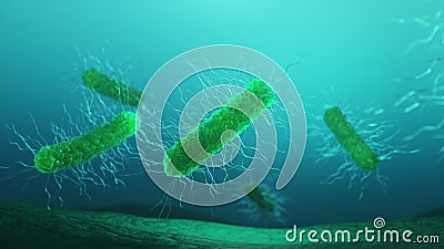


Bacteria Escherichia Coli Stock Footage Video Of Cell


Biol 230 Lab Manual Lab 1
E Coli Under The Microscope Types Techniques Gram Stain Bacteria Under Microscope Png Bacteria Under Microscope 1000x Share this post 0 Response to "Bacteria Under Microscope 1000x" Post a Comment Newer Post Older Post Home Subscribe to Post Comments (Atom) Iklan Atas ArtikelMany bacteria look like E coli when examined under the microscope (if not stained Enterobacteriaceae, Bacillus, cornyeformeImage taken at 1000x oil magnification and contributed by the Kansas Department of Health and Environment Trophozoite of E histolytica with ingested erythrocytes (arrow), under differential interference contrast (DIC) microscopy (Entamoeba coli, E hartmanni, E gingivalis, Endolimax nana,


Gram Stain
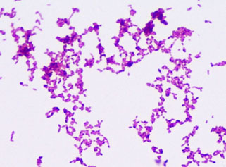


Bacterial Staining Microbiology Images Photographs And Videos Of Gram Acid Fast Endospore
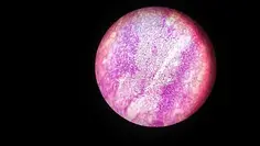


E Coli Under The Microscope Types Techniques Gram Stain Hanging Drop Method
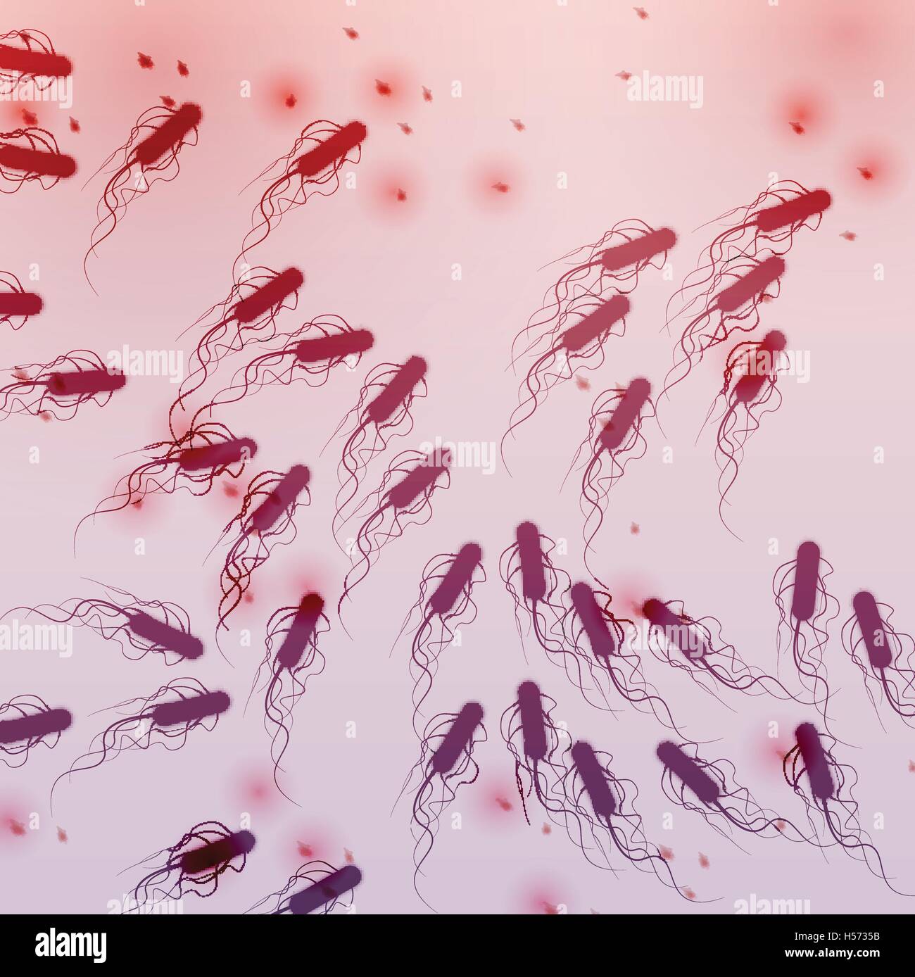


Bacillus Subtilis High Resolution Stock Photography And Images Alamy


Pin On Microscopic Organisms


What Does An E Coli Bacteria Look Like Under A Microscope Quora


What Does An E Coli Bacteria Look Like Under A Microscope Quora


Bacterial Microscopy Streaked Images


3 Microscopic View 1000x Of Escherichia Coli Download Scientific Diagram
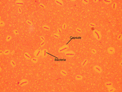


Micromorphology Slides Microbiology Resource Center Truckee Meadows Community College


Is A 1 000x Zoom On A Microscope Enough To See Bacteria Cells Quora


Pathogenic E Coli


Pbstatemicrobiology Licensed For Non Commercial Use Only Gram Stain


Gram Positive And Gram Negative Rods Microscopy Microbiology Microbiology Lab
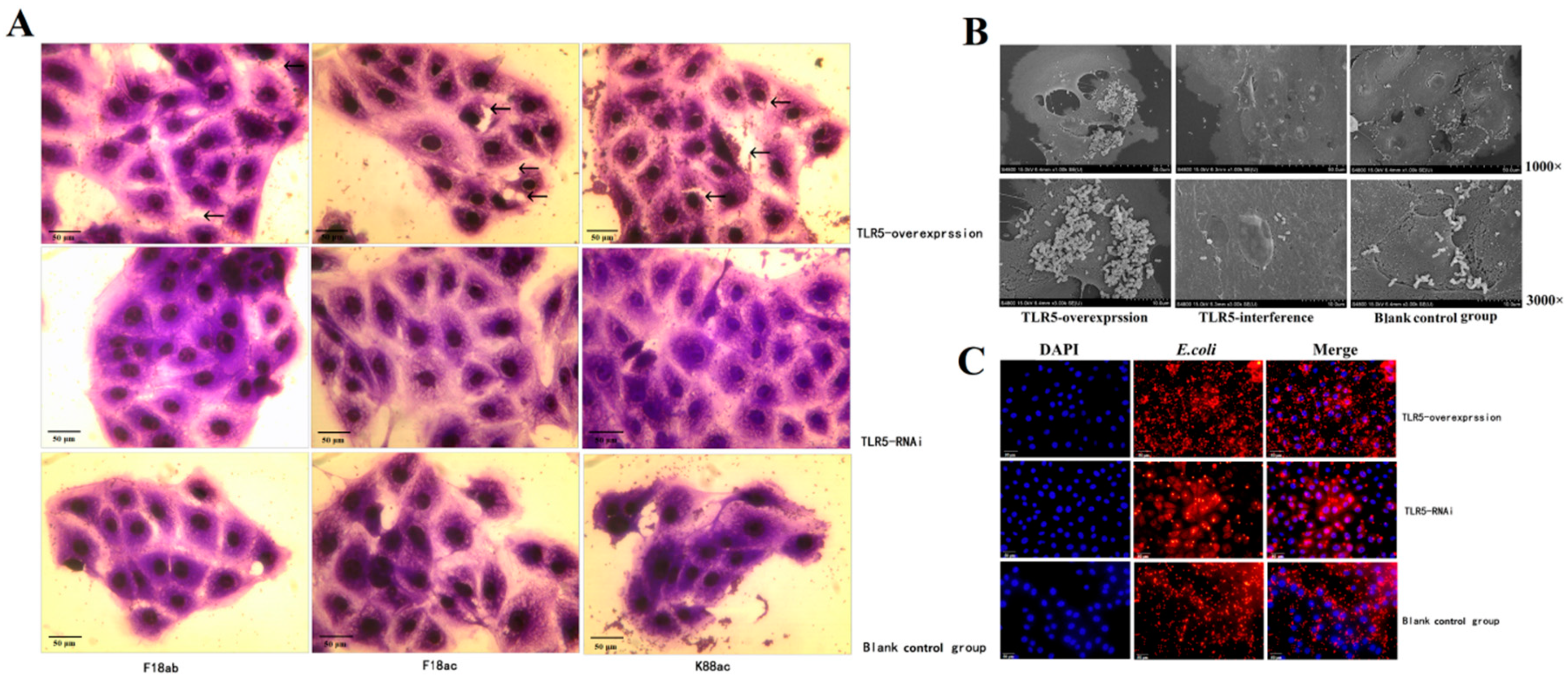


Animals Free Full Text Regulation And Molecular Mechanism Of Tlr5 On Resistance To Escherichia Coli F18 In Weaned Piglets Html
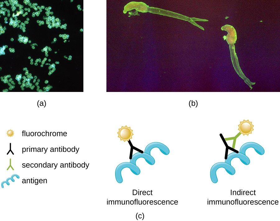


Instruments Of Microscopy Microbiology


Q Tbn And9gcqy Atzkf0po8c8x1yoohusnpjlpu6pnvquhzmi5dmxlldlhkom Usqp Cau


Endospore Staining Principle Procedure And Results Learn Microbiology Online
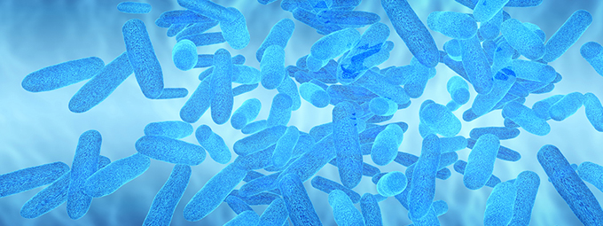


What Magnification Do I Need To See Bacteria Westlab
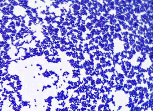


Bacterial Staining Microbiology Images Photographs And Videos Of Gram Acid Fast Endospore
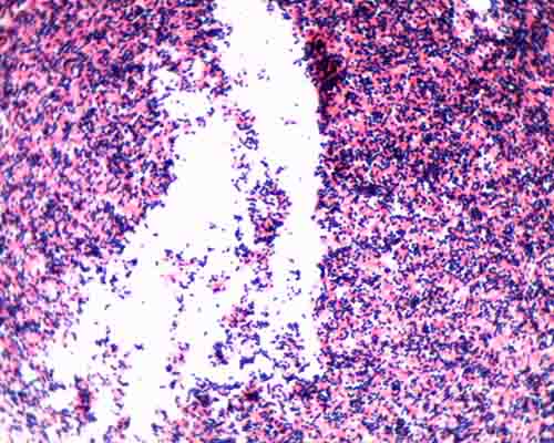


Gram Stain Microbiology Images Photographs


Lab 1


Www Mccc Edu Hilkerd Documents Bio1lab3 Exp 4 Pdf


Structure And Function Of Bacterial Cells


File E Coli At x Original Jpg Wikipedia


How Bacillus Spores Look Like Under Light Microscope


Microscopy Aquarium Advice Aquarium Forum Community


What Does An E Coli Bacteria Look Like Under A Microscope Quora



Escherichia Coli Bacteria E Coli Stock Footage Video 100 Royalty Free Shutterstock
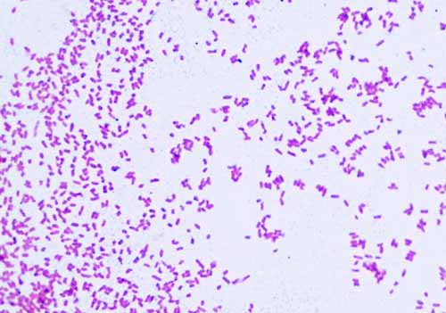


Gram Negative Bacteria Images Photos Of Escherichia Coli Salmonella Enterobacter
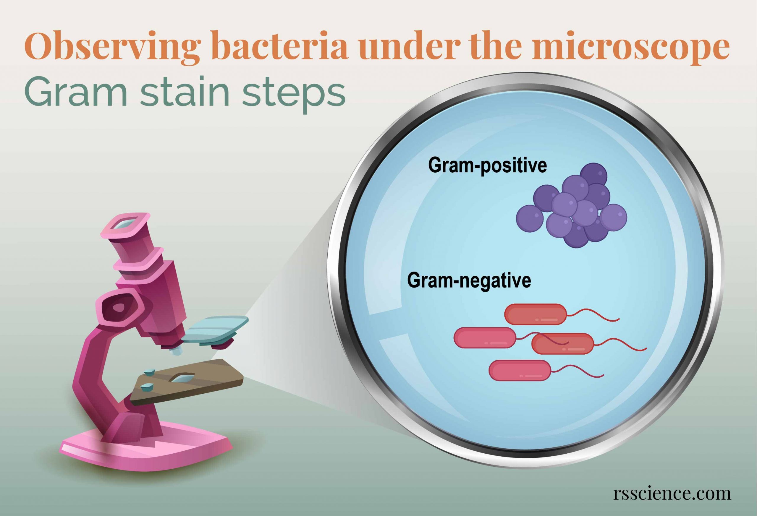


Observing Bacteria Under The Microscope Gram Stain Steps Rs Science


Gram Stain
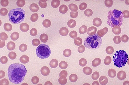


How These 26 Things Look Like Under The Microscope With Diagrams
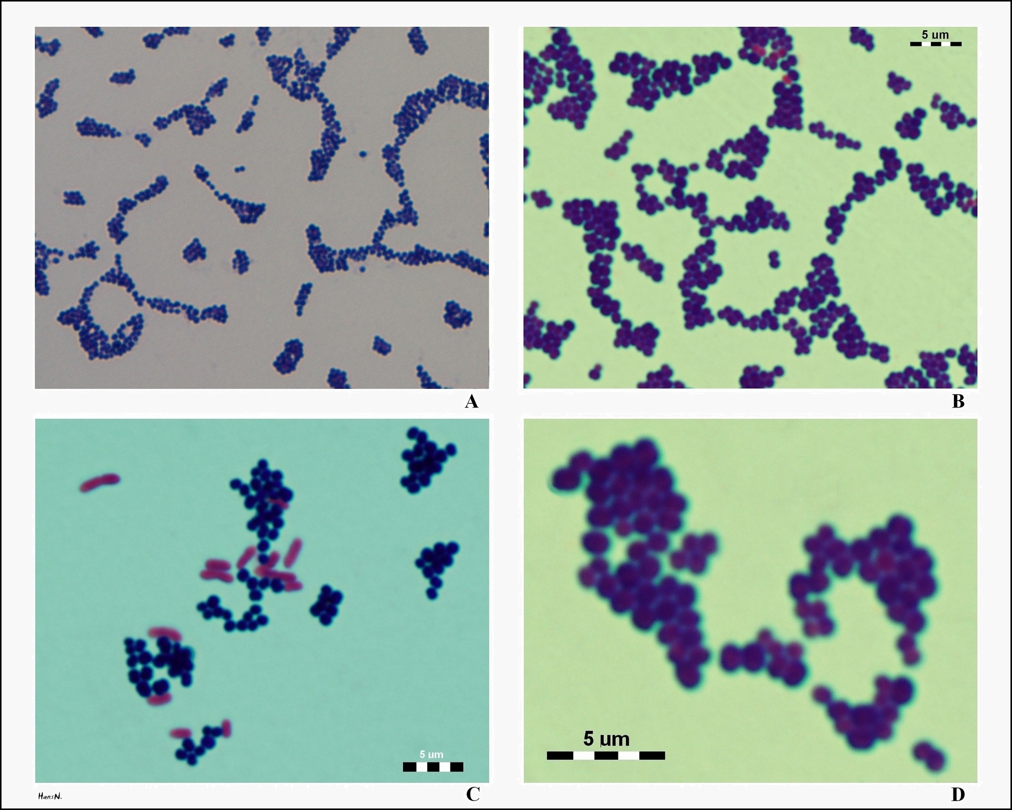


S Aureus Under The Microscope Microscopic Appearance And Morphology Of S Aureus Cell Arrangement


Bacteria Escherichia Coli Stock Footage Video Of Cell


Direct E Coli Cell Count At Od600 Tip Biosystems


Coli Stock Video Footage Royalty Free Coli Videos Pond5


Effect Of The Compound No On Cell Morphology Of E Coli Cells Download Scientific Diagram


Ecoli Under Microscope X1000 Youtube


Biol 230 Lab Manual Lab 1


Q Tbn And9gcswouuht13c4cxzkdgvwicdnmwhgkfjwlh40a Eerzzlpwjtmyt Usqp Cau


How Microbes Grow Science In The News


Figure 3 From Avirulent Gram Negative Bacteria E Coli K 12 Or E Coli C Compared With Gram Positive Virulent Diplococcic Streptoccocus Pneumonia Semantic Scholar


Bacteria Streaked Images


Microscope Imaging Of Methylene Blue Stained E Coli Cells Harbouring Download Scientific Diagram


Http Coltonanderson1 Weebly Com Uploads 2 4 3 0 Manual Pdf


Microscopy For The Winery Viticulture And Enology


Biol 230 Lab Manual Lab 1


Q Tbn And9gcqkye60ou Johpr02n Mbv1fferrjpdh Lnct7ymdf5qhyia1ld Usqp Cau


Pathogenic E Coli


Micromorphology Slides Microbiology Resource Center Truckee Meadows Community College


E Coli Under The Microscope Types Techniques Gram Stain Hanging Drop Method


Asmscience Examination Of Gram Stains Of Urine


Solved In This Section You Will View A Random Set Of Micr Chegg Com


Observing Bacteria Under The Light Microscope Microbehunter Microscopy


Escherichia Coli Bacteria E Coli Stock Footage Video 100 Royalty Free Shutterstock


Bacteria Escherichia Coli Stock Footage Video Of Cell
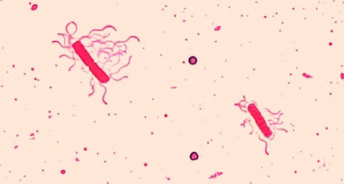


2 4 Staining Microscopic Specimens Microbiology Canadian Edition


Q Tbn And9gcsfopn62iwqhtvgb1rq4d6kbsyd6o90fpwmgbqlwwfpirnq5qmi Usqp Cau


Biol 230 Lab Manual Lab 1
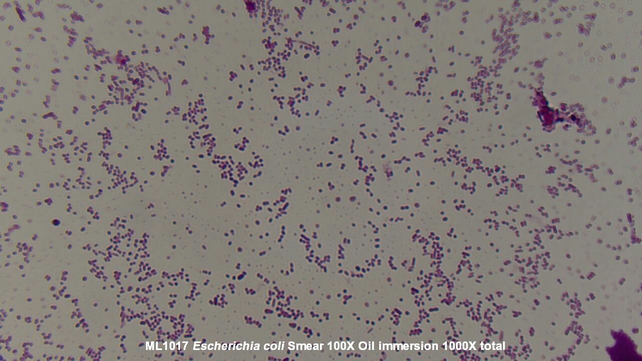


Slide Escherichia Coli


56 Questions With Answers In Gram Staining Scientific Method
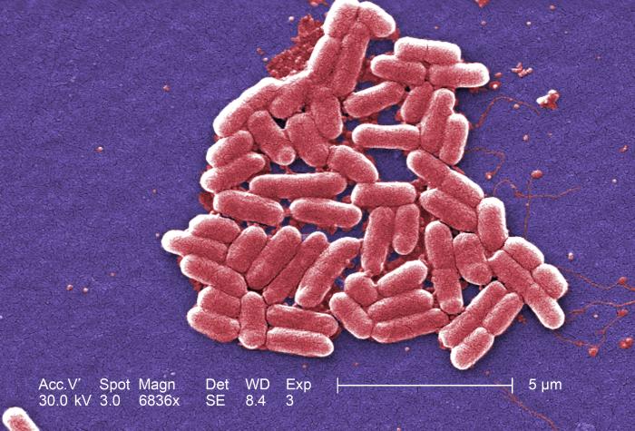


Details Public Health Image Library Phil


Escherichia Coli Bacteria E Coli Stock Footage Video 100 Royalty Free Shutterstock


Www Mccc Edu Hilkerd Documents Bio1lab3 Exp 4 Pdf


Whats Your Microscopy Setup Advanced Mycology Shroomery Message Board
.jpg)


Escherichia Coli 400x Escherichia Coli 400x Manufacturers Escherichia Coli 400x Suppliers Escherichia Coli 400x Exporters Escherichia Coli 400x In India


Lab 1


Observing Bacteria Under The Light Microscope Microbehunter Microscopy


What Does An E Coli Bacteria Look Like Under A Microscope Quora
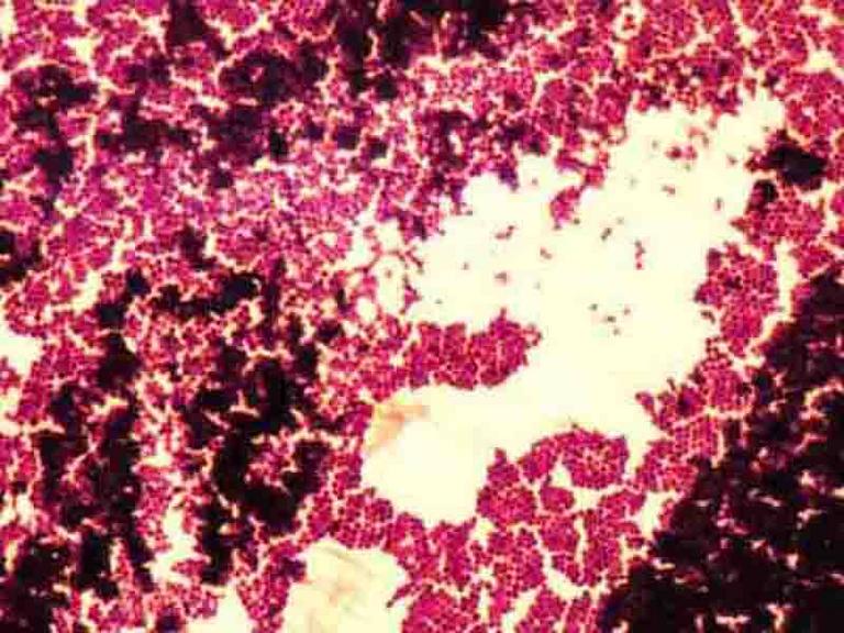


Bacterial Staining Microbiology Images Photographs And Videos Of Gram Acid Fast Endospore


27 Earth Bacteria E Coli


コメント
コメントを投稿